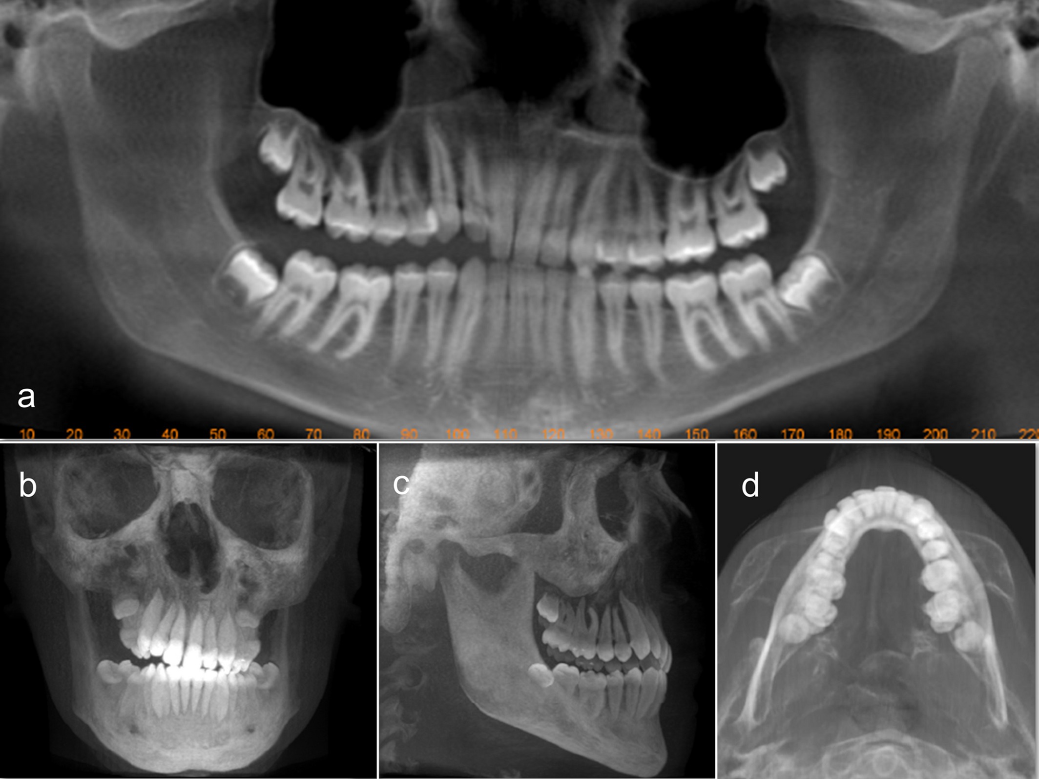
Ever wonder about the radiation you're exposed to during dental procedures, especially with those fancy 3D scans? Well, you're in the right place to learn all about it. In this article, we're diving into the world of CBCT (Cone Beam Computed Tomography) scans – a game-changer in the field of dental and medical imaging.
From understanding how much radiation in a CBCT scan you're actually exposed to, to exploring measures on mitigating radiation risks, we've got you covered. Stay with us as we unveil practical insights on CBCT dental scan radiation and delve into the specifics of molar CBCT radiation dosage. Your health and safety are paramount, and knowledge is your first line of defense.
Understanding CBCT: A Quick Overview of Radiation Dosage
When we talk about molar CBCT radiation dosage, it's essential to break down what that really means for you, the patient.

Cone Beam Computed Tomography (CBCT) has revolutionized dental imaging by providing 3D images that offer unparalleled diagnostic clarity. However, this advanced technology raises valid concerns about radiation exposure. The dosage from a molar CBCT scan generally falls within the range considered safe for medical diagnostic procedures.
The key to understanding this lies in comparing it to traditional dental X-rays. While CBCT scans offer a higher level of detail, they do so at a radiation dose that is only marginally higher than conventional dental radiographs when adjusted for diagnostic value. This nuanced perspective is crucial for making informed decisions about your dental care, emphasizing the balance between innovative diagnostic techniques and radiation safety.
How Much Radiation in a CBCT Scan? – Demystifying the Numbers
The question of how much radiation in a CBCT scan is frequently asked, embodying concerns about exposure during dental imaging.
It's vital to understand that radiation in CBCT scans is measured in millisieverts (mSv), providing a way to quantify exposure. A typical CBCT scan can range from 0.01 mSv to 0.3 mSv depending on the specifics of the scan, such as the field of view and the resolution required.
To put this into perspective, it's less than the yearly background radiation received from natural sources, and only a fraction of what you would be exposed to during a medical chest CT scan, which can be over 7 mSv. This comparison not only demystifies the amount of radiation but also contextualizes it within the broader spectrum of everyday and medical radiation exposure.
CBCT Dental Scan Radiation: What Patients Need to Know
Understanding CBCT dental scan radiation is paramount for patients contemplating undergoing this type of imaging.
The radiation emitted during a CBCT scan is adapted to achieve high-quality images with the least amount of exposure necessary. It's a principle known as ALARA (As Low As Reasonably Achievable).

For dental scans, particularly, this means the radiation is finely tuned to encompass only the area of interest — say, a single molar or the entire jaw — thereby limiting the patient's overall exposure.
Knowing this, patients can have peace of mind that CBCT technology is designed with their safety in mind, utilizing the minimal radiation necessary to achieve critical diagnostic images. This reassurance is crucial in dissipating the fog of apprehension that often surrounds the topic of radiation.
Measuring Radiation in CBCT Scans: Techniques and Tools
The measuring of radiation in CBCT scans is an exact science, utilizing sophisticated techniques and tools to ensure precision and safety.
Dosimeters are devices used to measure the absorbed radiation dose during the scan. Their usage provides a direct reading of the patient's exposure, enabling practitioners to adhere strictly to the ALARA principle.
Furthermore, recent advancements in CBCT technology have introduced software algorithms capable of optimizing scan parameters in real-time, effectively minimizing unnecessary radiation while upholding the quality of the diagnostic image. This ongoing evolution in measuring and managing radiation exposure showcases the dental industry's commitment to patient safety without compromising the diagnostic value of CBCT scans.
Mitigating CBCT Scan Radiation: Effective Strategies for Safety
Mitigating CBCT scan radiation involves a multi-pronged approach to ensure patient safety remains paramount.

One effective strategy is the utilization of protective gear, such as lead aprons and thyroid collars, which can significantly reduce radiation exposure to vulnerable areas of the body. Additionally, dental professionals are increasingly relying on digital imaging software that enhances the clarity and utility of CBCT images, allowing for lower radiation doses during scans.
Training for practitioners in radiation management and the latest CBCT technologies also forms a critical line of defense, ensuring that every scan is performed with the utmost care for radiation minimization. These strategies, when combined, form a robust framework for safeguarding patients, marking a significant stride in responsible radiographic imaging.
Comparing Radiation: CBCT Scans Vs. Traditional Imaging
Comparing the radiation from CBCT scans to traditional imaging methods sheds light on the advancements and safety improvements in dental diagnostics.
Traditional imaging techniques, such as panoramic x-rays, are known for their lower radiation exposure but at the expense of comprehensive diagnostic information. CBCT scans, while involving slightly higher radiation doses, provide exhaustive 3D images that offer critical insights impossible to garner from 2D imaging.
This added dimension is particularly crucial for complex cases requiring detailed anatomical information. The perspective gained from this comparison underscores the importance of selecting the appropriate imaging technique based on the diagnostic needs, balancing radiation exposure with the necessity of obtaining high-fidelity images that can inform better clinical decisions.

